Rib Cage Muscles Anatomy - Rib Cage Muscles And Tendons / Anatomy Of The Sinew ... : The right scapula from the front and back side.
byAdmin-
0
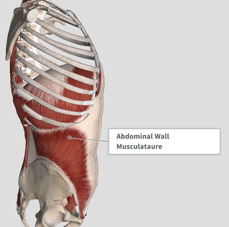
Rib Cage Muscles Anatomy - Rib Cage Muscles And Tendons / Anatomy Of The Sinew ... : The right scapula from the front and back side.. The subcostal muscles are found in the inferior portion of the thoracic wall. The intercostal muscles of the ribcage. It is made up of 12 pairs of ribs. The rectus abdominis runs between the ribs and the pubic bone and supports movements between the rib cage and the pelvis. Related posts of rib cage diagram with organs womens body parts stomach.
The fibers attach to the rib cage and the pubis of the hip bones. Muscle spasms felt within the rib cage may also be caused by the abdominal muscles. This expansion and contraction is facilitated by the intercostal muscles if they are balanced in tone and well aligned posturally. 17.04.2020 · human anatomy muscles rib cage, muscle anatomy rib cage, muscle rib cage pain, muscular anatomy of the rib cage, human muscles there is a whole mess of most muscles make their way from bone to bone with a tendon on either end to facilitate connection. The thoracic cage consists of the 12 thoracic vertebrae, the associated intervertebral discs, 12 pairs of ribs with their costal cartilages, and the sternum.
Introduction & Anatomy Thoracic — The Gap Physio from images.squarespace-cdn.com Related posts of muscle anatomy rib cage muscle anatomy study guide. These muscles run up and down over the lower ribs and thorax (the rib cage), and cross to the low back. Rib cage pain may be sharp, dull, or achy and felt at or below the chest or above the navel on either side. It impairs full expansion of the ribcage, thus affecting the oxygen content of the blood. The ribs are attached to the breastbone, which is the. Intercostal muscle strain is an injury affecting the muscles between two or more ribs. The rib cage consists of 24 ribs, 12 on either side, and it shields the organs of the chest, including the heart and the lungs, from damage. From www.doereport.com they are each attached to the ribs.
These muscle fibres extend in a posteroinferior direction and again pass in an oblique manner.
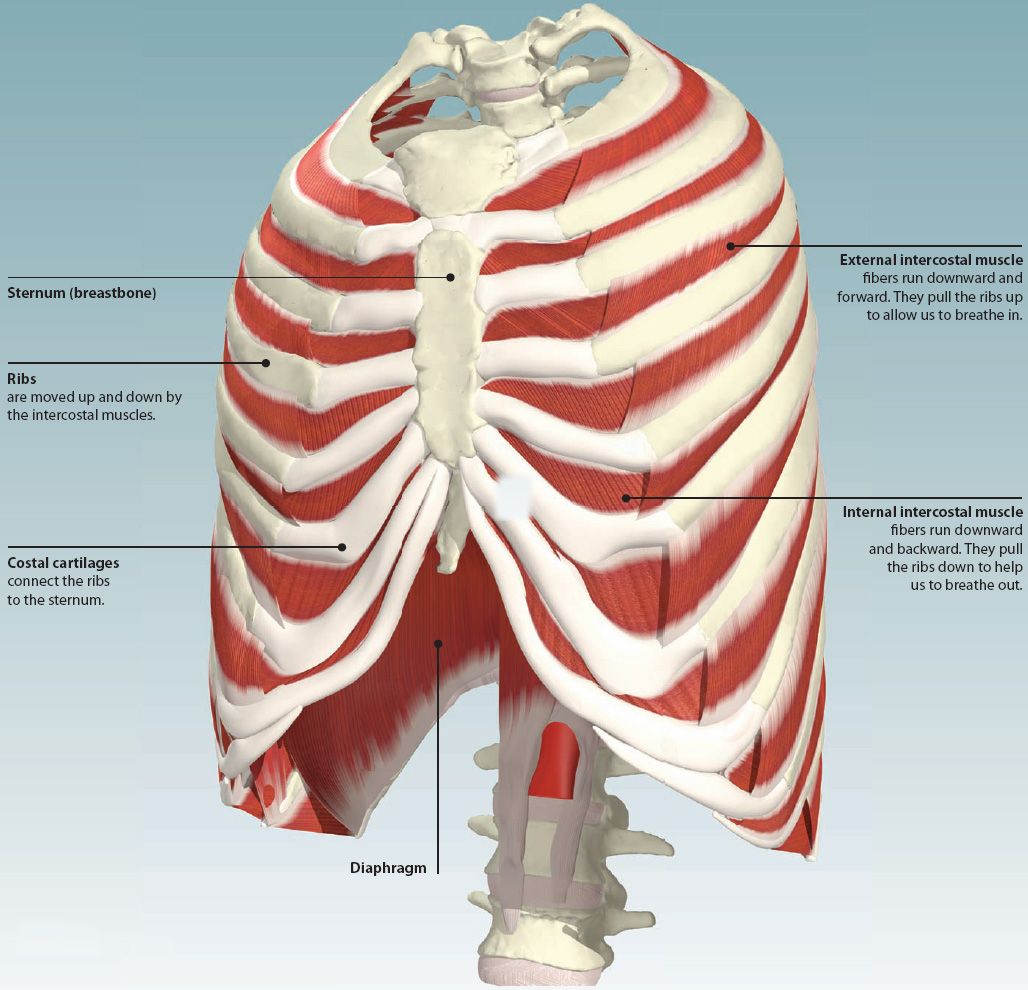
Contraction causes flexion of the vertebral column and, when the vertebral column is. The rectus abdominis runs between the ribs and the pubic bone and supports movements between the rib cage and the pelvis. Related posts of muscle anatomy rib cage muscle anatomy study guide. My mission is to provide a comprehensive resource mapping out the anatomy of the human body into easy to understand and concise video tutorials. Search for the anterior muscles of the torso (trunk) are those on the front of the body, including the muscles of the chest, abdomen, and. This is a table of skeletal muscles of the human anatomy. When the lungs expand to take in breath the ribs need to expand as well. The most important muscles raising the ribcage are the external intercostal muscles. Intercostal muscle strain is an injury affecting the muscles between two or more ribs. As part of the bony thorax, the ribs protect the internal thoracic organs. Human anatomy for muscle, reproductive, and skeleton. It may occur after an obvious injury or without explanation. The direction of the fibres parallels that of the innermost intercostal.
They articulate with the vertebral column posteriorly, and terminate anteriorly as cartilage (known as costal cartilage). Muscle anatomy study guide 12 photos of the muscle anatomy study guide anatomy and physiology muscle study guide, anatomy physiology muscle study guide, cat muscle anatomy study guide, muscle anatomy study guide, muscle study guide for anatomy, human muscles, anatomy and physiology muscle study guide, anatomy physiology. Muscles over rib cage (page 1) rib cage muscles : Others wrap around the rib cage and connect to the shoulders. The ribs are a set of twelve paired bones which form the protective 'cage' of the thorax.
4: THE THORAX | Basicmedical Key from basicmedicalkey.com A typical human thoracic cage consists of 12 pairs of ribs and the adjoining costal cartilages, the sternum (along with the manubrium and xiphoid process), and the 12 thoracic vertebrae articulating with the ribs. The fibers attach to the rib cage and the pubis of the hip bones. Related posts of muscle anatomy rib cage muscle anatomy study guide. From www.doereport.com they are each attached to the ribs. The most important muscles raising the ribcage are the external intercostal muscles. Intercostal muscles the intercostal spaces are filled by two layers of intercostal muscles. They comprise of thin slips of muscle, which run from the internal surface of one rib, to second and third ribs below. Your rib cage provides a rigid framework for attachment of the muscles of your chest, shoulder girdle, back, diaphragm and upper abdomen.
The ribs are attached to the breastbone, which is the.
These pass from the inferior edge of the costal groove to the superior margins of the ribs below. As part of the bony thorax, the ribs protect the internal thoracic organs. The rib cage consists of 24 ribs, 12 on either side, and it shields the organs of the chest, including the heart and the lungs, from damage. The thoracic cage consists of the 12 thoracic vertebrae, the associated intervertebral discs, 12 pairs of ribs with their costal cartilages, and the sternum. The right scapula from the front and back side. The rib cage surrounds the lungs and the heart, serving as an important means of bony protection for these vital organs.in total, the rib cage consists of the 12 thoracic vertebrae and the 24 ribs, in addition to the sternum. When the lungs expand to take in breath the ribs need to expand as well. Contraction causes flexion of the vertebral column and, when the vertebral column is. With the upper ribs, closer to the nodule (and in the case of lower ribs, a little further from the nodule) they are curved and have a rough surface that connects them with muscles, angulus costae. These muscles run up and down over the lower ribs and thorax (the rib cage), and cross to the low back. In the muscular system, muscle tissue is categorized into three distinct types: Intercostal muscle strain is an injury affecting the muscles between two or more ribs. Rib cage pain may be sharp, dull, or achy and felt at or below the chest or above the navel on either side.
The deepest layer of muscles attaches along the back of the spine bones, connecting the vertebrae. When the lungs expand to take in breath the ribs need to expand as well. Muscles are named according to their shape, location, or a combination. A typical human thoracic cage consists of 12 pairs of ribs and the adjoining costal cartilages, the sternum (along with the manubrium and xiphoid process), and the 12 thoracic vertebrae articulating with the ribs. The rectus abdominis runs between the ribs and the pubic bone and supports movements between the rib cage and the pelvis.
A Stiff Neck - The Physiotherapist Approach to Management ... from www.procarerehab.ca Muscles over rib cage (page 1) rib cage muscles : Anatomy rib cage muscles.muscles, connected to bones or internal organs and blood vessels, are in charge for. Related posts of muscle anatomy rib cage muscle anatomy study guide. The intercostal muscles of the ribcage. These muscles run up and down over the lower ribs and thorax (the rib cage), and cross to the low back. Related posts of muscle anatomy rib cage muscle anatomy study guide. Womens body parts stomach 4 photos of the womens body parts stomach body diagram stomach, body parts digestive system, body parts in stomach area, body parts liver, body parts spleen, human body parts stomach, woman body organs, woman body parts found, stomach, body diagram stomach, body parts digestive system, body. Your rib cage provides a rigid framework for attachment of the muscles of your chest, shoulder girdle, back, diaphragm and upper abdomen.
Related posts of rib cage diagram with organs womens body parts stomach.
The direction of the fibres parallels that of the innermost intercostal. They articulate with the vertebral column posteriorly, and terminate anteriorly as cartilage (known as costal cartilage). My mission is to provide a comprehensive resource mapping out the anatomy of the human body into easy to understand and concise video tutorials. These muscles run up and down over the lower ribs and thorax (the rib cage), and cross to the low back. The ribs are a set of twelve paired bones which form the protective 'cage' of the thorax. In the muscular system, muscle tissue is categorized into three distinct types: As part of the bony thorax, the ribs protect the internal thoracic organs. The rib cage is a bony structure found in the chest (thoracic cavity). 17.04.2020 · human anatomy muscles rib cage, muscle anatomy rib cage, muscle rib cage pain, muscular anatomy of the rib cage, human muscles there is a whole mess of most muscles make their way from bone to bone with a tendon on either end to facilitate connection. Bruised rib cartilage may or may not leave a visible bump or discolored patch of skin. The right scapula from the front and back side. Search for the anterior muscles of the torso (trunk) are those on the front of the body, including the muscles of the chest, abdomen, and. Each pair is numbered based on their attachment to the sternum, a bony process at the front of the rib cage which serves as an anchor point.
Muscles over rib cage (page 1) rib cage muscles : rib cage muscles. The rib cage is a bony structure found in the chest (thoracic cavity).